Formation of The Lungs
Lung development is characterized by periods of development, and
develops in a proximal-distal direction, beginning with the largest bronchi and
proceeding outward. This means that lung development is heterogeneous, as the
proximal pulmonary tissue will be in a more advanced period of development that
the distal pulmonary tissue.
1. Pseudoglandular period (weeks 7-16). The developing lung resembles an exocrine gland, hence the name. The numerous endodermal tubules are lined by simple columnar epithelium. Each endodermal tube branches into 15 to 25 terminal bronchioles. Respiration is not possible during this period, and premature infants cannot survive.
2. Canalicular period (weeks 16-24). The terminal bronchioles branch into three or more respiratory bronchioles, which in turn branch into three to six alveolar ducts. The terminal bronchioles, respiratory bronchioles, and alveolar ducts are now lined by a simple cuboidal epithelium. Premature infants born before week 20 rarely survive.
2. Canalicular period (weeks 16-24). The terminal bronchioles branch into three or more respiratory bronchioles, which in turn branch into three to six alveolar ducts. The terminal bronchioles, respiratory bronchioles, and alveolar ducts are now lined by a simple cuboidal epithelium. Premature infants born before week 20 rarely survive.
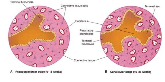
Figure 3. Early stages of lung development. (Moore, K, and Persaud, T. 2008)
3. Terminal Sac period (weeks 24-birth). The alveolar ducts bud off terminal sacs, which dilate and expand into the surrounding mesoderm. The terminal sacs are separated from each other by primary septa. The simple cuboidal epithelium within the terminal sacs differentiates into Type 1 and Type 2 alveolar epithelial cells. Type 1 alveolar epithelial cells are thin flat cells that make up part of the blood-air barrier. Type 2 alveolar epithelial produce surfactant, which is a fluid capable of lowering surface tension at the air-alveolar interface.
The terminal sacs are surrounded by mesoderm containing a rapidly proliferating capillary network. The capillaries make intimate contact with the terminal sacs, and establish a blood-air barrier with the Type 1 alveolar epithelial cells. Premature infants born between week 25 and 28 can survive with intensive care; this is dependent on the surfactant levels and vascularization
4. Alveolar Period (birth- 8 years of age). The terminal sacs are partitioned by secondary septa to form adult alveoli. 20-70 million alveoli are present at birth, and increase to about 300-400 million alveoli by 8 years of age. This increase of alveoli is mainly due to the formation of secondary septa that partition existing alveoli. The increase in the size of the lungs after birth is due to an increase in the number of respiratory bronchioles.
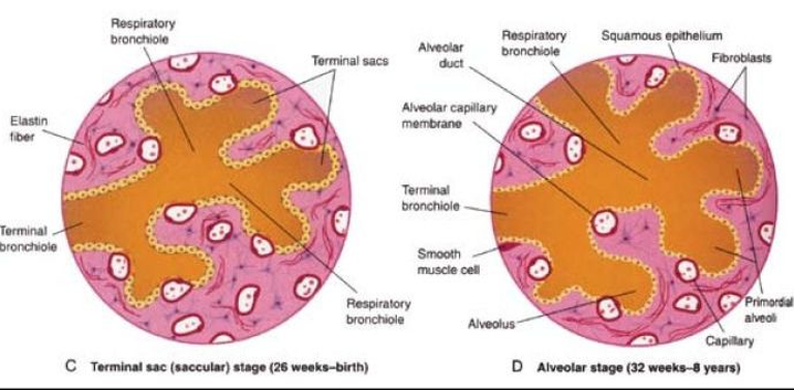
Figure 4. Later stages of lung development. Note the bulging of the capillaries into the alveoli and terminal sacs. (Moore, K, and Persaud, T. 2008)
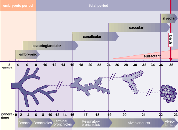
Figure 5. An overview of the periods of lung development. http://www.embryology.ch/anglais/rrespiratory/phasen02.html. Downloaded on April 8, 2011.
