Formation of The Trachea and Bronchi
The
tracheal epithelium and glands are derived from endoderm. The tracheal smooth
muscle, connective tissue, and C-shaped cartilage rings are derived from
visceral mesoderm. The lung bud divides into two bronchial buds.The lung buds expand throughout a part of coelomic cavity called the pericardioperitoneal canal. This canal will be separated from the peritoneal and pericardial cavity by the pleuroperitoneal and pleuropericardial membranes, respectively, thus forming the primitive pleural cavities. During further development the main bronchi divide approximately 17 times, forming smaller canals (canalicular phase).
In week 5 of development, bronchial buds enlarge to form main (primary) bronchi. The right main bronchus is larger and more vertical than the left main bronchus, and this remains throughout adult life and is the reason why there’s a greater chance of foreign bodies lodging on the right side than on the left. The main bronchi then subdivide into lobar (secondary bronchi); 3 on the right side and two on the left. The lobar bronchi further subdivide into segmental (tertiary) bronchi; 10 on the right and 9 on the left, which further subdivide into subsegmental bronchi (see Figure 2). The segmental (tertiary) bronchus gives rise to the bronchopulmonary segments, which are segments of lung tissue that are morphologically and functionally separate respiratory units of the lung.
As the bronchi develop, they expand laterally and caudally into a space known as the primitive pleural cavity. The visceral mesoderm covering the outside of the bronchi develops into visceral pleura, and somatic mesoderm covering the inside of the body wall develops into parietal pleura. The space between the visceral and parietal pleura is called the pleural cavity.
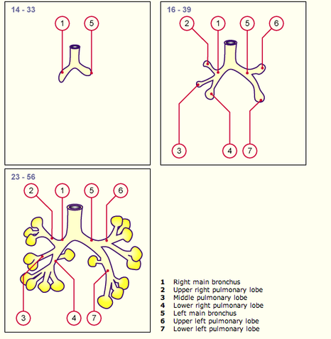
Figure 2. Shows the divisions of the bronchi. http://www.embryology.ch/anglais/rrespiratory/phasen02.html Downloaded on April 8, 2011
