Formation of The Diaphragm
The diaphragm, which separates the
abdominal cavity from the plerual cavities, is a composite structure. Development begins during the third week of gestation and is complete by the eighth week. The embryonic structures from which the diaphragm develops include the (1) septum transversum,
the (2) dorsal esophageal mesentery, the (3) pleuroperitoneal folds, and (4)
body wall mesoderm. Pleuroperitoneal folds arise from the posterior body wall
and lie in a plane that is parallel to the septum transversum and perpendicular
to the pleuropericardial folds. Failure in the development of the pleuroperitoneal folds and subsequent muscle migration results in congenital defects.
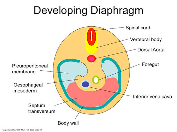
Figure 6. The embryological origins of the diaphragm. http://embryology.med.unsw.edu.au/Notes/respire7.html. Downloaded on April 8, 2011.
The diaphragm is innervated from the third through fifth cervical spinal cord segments via the phrenic nerve. The diaphragm develops initially in a cephalic region and during development (anteroposterior folding of the embryo) descends into a more caudal thoracic position. The phrenic nerves travel with the diaphragm and come to lie within the fibrous pericardium.
